Many people may face severe abdominal pain without realizing it could be due to gallstones or gallbladder inflammation. Gallstones, hardened deposits in the gallbladder, can block bile ducts and lead to acute cholecystitis, a painful condition requiring prompt medical care. Knowing the symptoms and available treatments is vital for managing these health issues.
This article provides information on recognizing symptoms, diagnosing the condition, and understanding the treatment options for gallbladder calculus and acute cholecystitis.
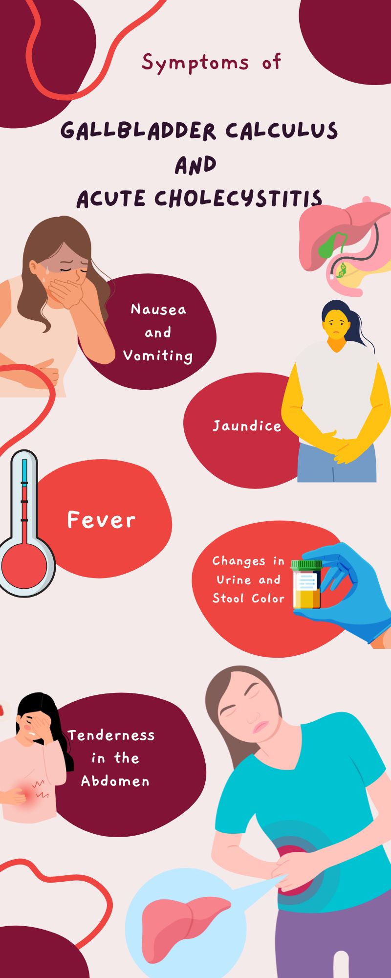
Symptoms of Gallbladder Calculus and Acute Cholecystitis
Pain is often the first and most noticeable sign of gallstones and gallbladder inflammation. Typically, this pain occurs in the upper right part of the abdomen and can be severe. It may radiate to the back or right shoulder. This pain usually comes on suddenly and can last for several hours.
Nausea and Vomiting
Many individuals experience nausea and vomiting alongside abdominal pain. These symptoms can be particularly intense after eating a large or fatty meal, as the gallbladder tries to release bile but is obstructed by gallstones.
Fever
A fever may accompany the other symptoms, indicating an infection in the gallbladder. The fever can be mild or high, depending on the severity of the inflammation and infection.
Tenderness in the Abdomen
Touching the abdomen, especially in the upper right quadrant, may cause tenderness. This tenderness is a sign of inflammation and should not be ignored.
Jaundice
In some cases, jaundice may occur, causing the skin and the whites of the eyes to turn yellow. This happens when gallstones block the bile ducts, preventing bile from being excreted from the liver and gallbladder.
Changes in Urine and Stool Color
Dark urine and pale stools can also be symptoms of blocked bile ducts. These changes occur because bile cannot reach the intestines to be excreted properly, affecting the color of waste products.
What Are the Main Causes?
Several factors contribute to the development of gallstones and acute cholecystitis. Understanding these causes and risk factors can help in both preventing and managing these conditions effectively.
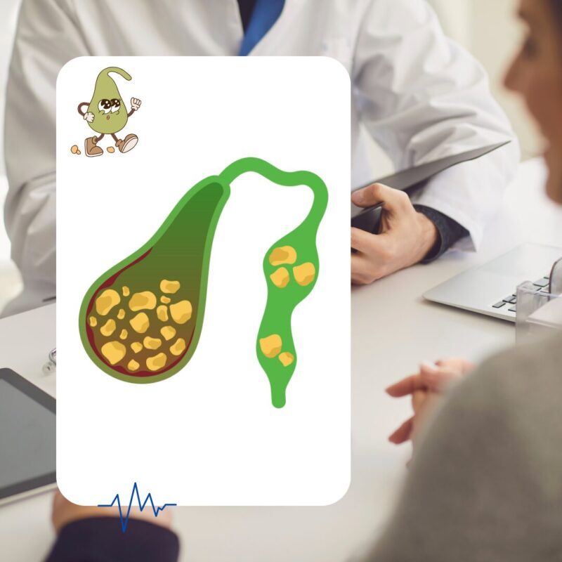
Gallstones Formation
Gallstones form when there is an imbalance in the substances that make up bile. Bile, produced by the liver, aids in digestion and is stored in the gallbladder. When the bile contains too much cholesterol, bilirubin, or not enough bile salts, it can harden into stones.
Factors that can contribute to this imbalance include:
- Excess Cholesterol: The liver may produce more cholesterol than the bile can dissolve, leading to stone formation. This is common in individuals who are obese or have a diet high in fat.
- Excess Bilirubin: Conditions such as liver cirrhosis, biliary tract infections, and certain blood disorders can cause the liver to produce too much bilirubin, leading to the formation of pigment stones.
- Insufficient Bile Salts: Reduced bile salt production can prevent the dissolution of cholesterol, leading to gallstones. This can be influenced by factors like rapid weight loss or fasting.
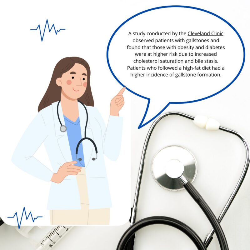
A study conducted by the Cleveland Clinic observed patients with gallstones and found that those with obesity and diabetes were at higher risk due to increased cholesterol saturation and bile stasis. Patients who followed a high-fat diet had a higher incidence of gallstone formation.
Bile Duct Blockage
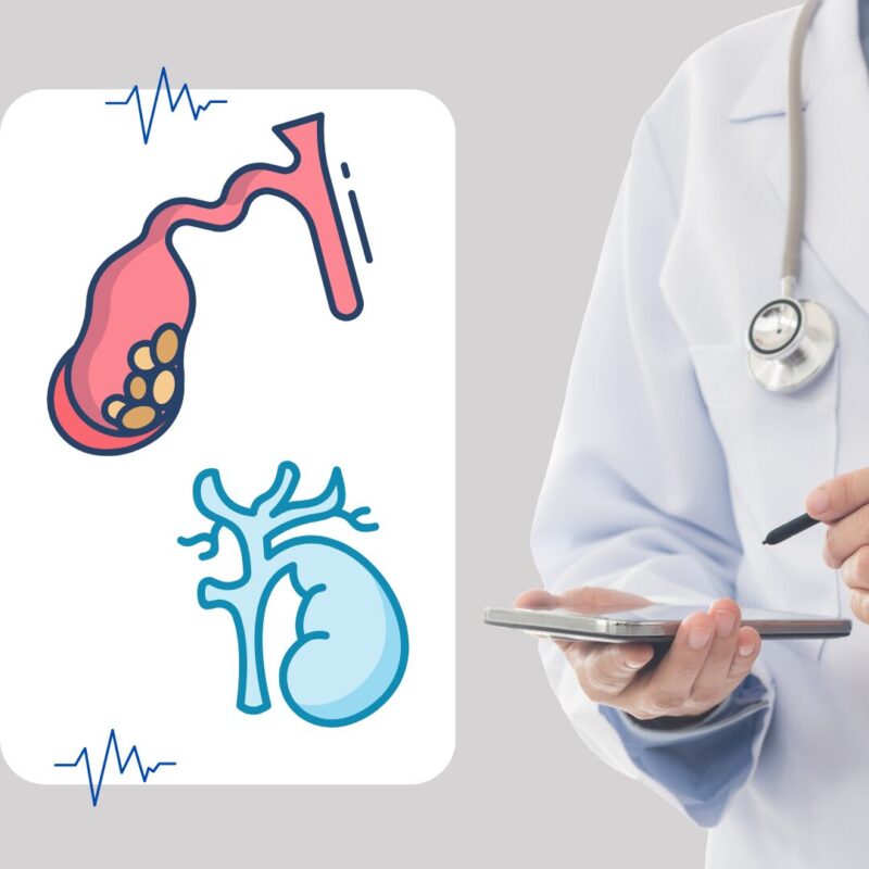
When gallstones block the cystic duct, bile builds up in the gallbladder, causing inflammation and infection, which is known as acute cholecystitis. This blockage can be temporary or persistent and is a major trigger for the condition.
Tumors and Bile Duct Strictures
Less commonly, tumors or strictures (narrowing of the bile ducts) can prevent the proper flow of bile, leading to its buildup and subsequent inflammation. These conditions require advanced imaging techniques for proper diagnosis and management.
Infections
Certain infections, including those caused by viruses such as HIV, can trigger gallbladder inflammation. These infections may cause a reaction that results in the formation of gallstones or the inflammation of the gallbladder.
Severe Illness
Critical illnesses, particularly those that result in sepsis or severe systemic infections, can decrease blood flow to the gallbladder, leading to inflammation. This is particularly seen in patients in intensive care units or those with severe trauma.
Genetic Factors
There is evidence that genetics play a role in the development of gallstones. Individuals with a family history of gallstones are more likely to develop them. Studies have shown that genetic variations can affect the composition of bile and the motility of the gallbladder.
Lifestyle and Dietary Factors
Lifestyle choices and diet also significantly influence the risk of developing gallstones. Diets high in cholesterol and low in fiber, sedentary lifestyle, and obesity are notable risk factors. Conversely, a diet rich in fruits, vegetables, and whole grains can reduce the risk.
Diagnostic
Diagnosing gallstones and acute cholecystitis requires a combination of clinical evaluation, laboratory tests, and imaging techniques to ensure accurate detection and effective treatment.
Clinical Evaluation
A thorough clinical history and physical examination are the first steps in diagnosing these conditions. Common symptoms include severe right upper abdominal pain, nausea, vomiting, and fever. A physical examination may reveal tenderness in the right upper quadrant and a positive Murphy’s sign (pain upon palpation under the rib cage during inhalation).
Laboratory Tests
Blood tests play a crucial role in the diagnosis. Elevated white blood cell counts indicate infection or inflammation. Liver function tests, including bilirubin, aspartate aminotransferase (AST), and alanine aminotransferase (ALT) levels, can indicate bile duct obstruction or liver involvement.
A study in the Journal of Clinical Medicine showed the importance of early laparoscopic cholecystectomy (ELC) for acute cholecystitis. It focused on using a combination of clinical assessments, lab tests, and imaging techniques to ensure accurate diagnosis.
Imaging Techniques
- Ultrasound (US): This is the most common and first-line imaging test for diagnosing gallstones and acute cholecystitis. Ultrasound is non-invasive, widely available, and cost-effective. It can detect gallstones, bile duct obstruction, and gallbladder wall thickening, which are indicative of acute cholecystitis. The sensitivity and specificity of ultrasound for acute cholecystitis are approximately 71% and 85%, respectively.
- Computed Tomography (CT) Scan: CT scans provide detailed images of the gallbladder and surrounding structures. They are particularly useful in complicated cases or when ultrasound results are inconclusive. CT scans can detect complications such as perforation, abscess formation, or pancreatitis.
- Hepatobiliary Iminodiacetic Acid (HIDA) Scan: This nuclear medicine test evaluates the function of the gallbladder and bile ducts. It involves injecting a radioactive tracer that is taken up by the liver and excreted into the bile. If the tracer fails to enter the gallbladder, it suggests a blockage, which is indicative of acute cholecystitis. HIDA scans have high sensitivity and specificity, often exceeding 90%, but their use is limited in emergency settings due to time and resource constraints.
- Magnetic Resonance Cholangiopancreatography (MRCP): This is a non-invasive MRI technique used to visualize the biliary and pancreatic ducts. MRCP is highly effective for detecting bile duct stones and other abnormalities without the need for contrast dye, making it safer for patients with kidney problems or allergies to contrast agents.
- Endoscopic Retrograde Cholangiopancreatography (ERCP): ERCP combines endoscopy and fluoroscopy to diagnose and treat conditions of the biliary or pancreatic ductal systems. It is particularly useful for removing bile duct stones and relieving obstructions. ERCP is both diagnostic and therapeutic but carries risks such as pancreatitis and infections.
Complications and Prevention
Understanding the potential complications of gallstones and acute cholecystitis, as well as effective prevention strategies, is essential for managing these conditions and minimizing risks.
Acute Cholecystitis
One of the most common complications of gallstones is acute cholecystitis, where the gallbladder becomes inflamed due to bile buildup. This can lead to severe pain, fever, and infection. If left untreated, it can result in more serious issues such as gangrene (death of gallbladder tissue) or a ruptured gallbladder, both of which are life-threatening and require immediate medical intervention.
Bile Duct Stones (Choledocholithiasis)
Gallstones can move from the gallbladder into the bile ducts, causing blockages that lead to jaundice (yellowing of the skin and eyes), pancreatitis (inflammation of the pancreas), or cholangitis (infection of the bile ducts). These conditions are serious and require prompt treatment, often involving endoscopic procedures to remove the stones and restore normal bile flow.
Pancreatitis
When gallstones block the ducts of the pancreas, it can cause pancreatitis, characterized by severe abdominal pain, nausea, vomiting, and elevated levels of pancreatic enzymes in the blood. This condition can range from mild to life-threatening and typically requires hospitalization.
Gallbladder Cancer
Although rare, chronic inflammation from gallstones can increase the risk of gallbladder cancer. This type of cancer is often diagnosed at an advanced stage due to its nonspecific symptoms and the difficulty in detecting it early.
Prevention
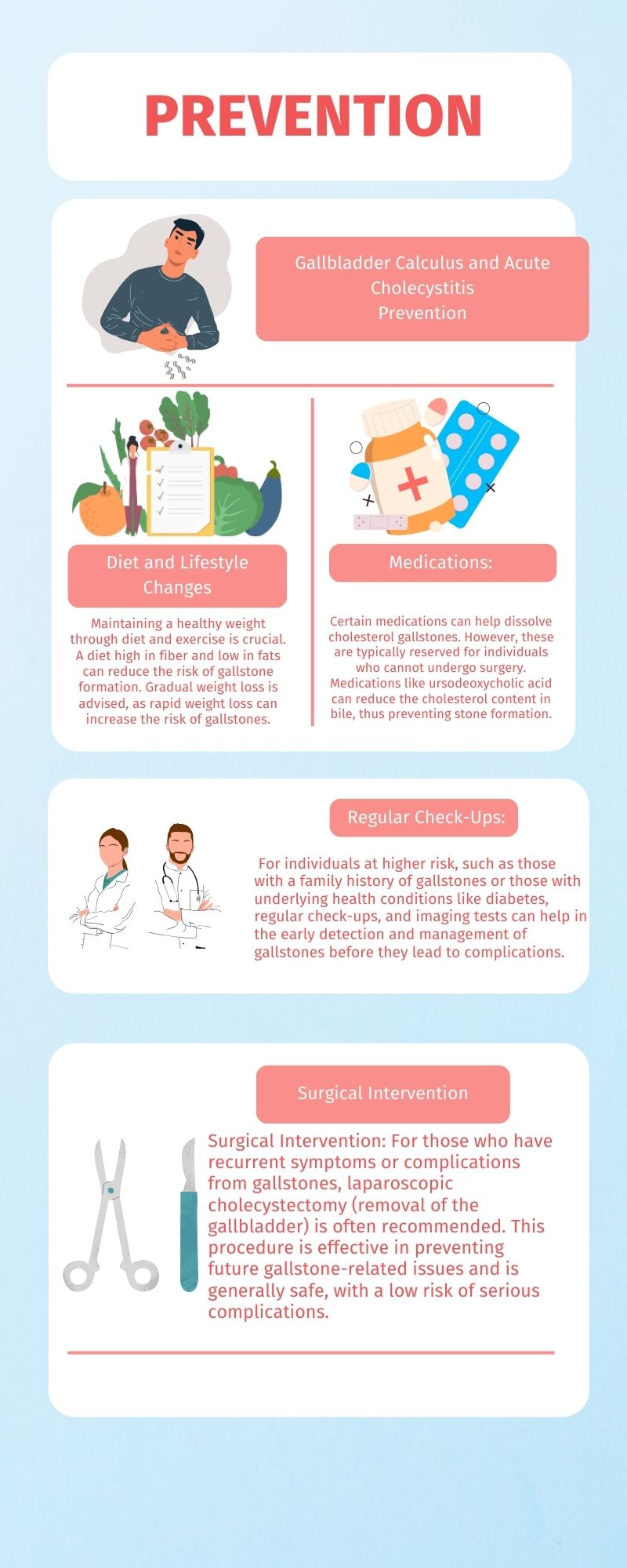
1. Diet and Lifestyle Changes: Maintaining a healthy weight through diet and exercise is crucial. A diet high in fiber and low in fats can reduce the risk of gallstone formation. Gradual weight loss is advised, as rapid weight loss can increase the risk of gallstones.
2. Medications: Certain medications can help dissolve cholesterol gallstones. However, these are typically reserved for individuals who cannot undergo surgery. Medications like ursodeoxycholic acid can reduce the cholesterol content in bile, thus preventing stone formation.
3. Regular Check-Ups: For individuals at higher risk, such as those with a family history of gallstones or those with underlying health conditions like diabetes, regular check-ups, and imaging tests can help in the early detection and management of gallstones before they lead to complications.
4. Surgical Intervention: For those who have recurrent symptoms or complications from gallstones, laparoscopic cholecystectomy (removal of the gallbladder) is often recommended. This procedure is effective in preventing future gallstone-related issues and is generally safe, with a low risk of serious complications.
Summary
Gallbladder calculus (gallstones) and acute cholecystitis are significant health concerns that require timely diagnosis and treatment to prevent severe complications. Understanding the symptoms, such as abdominal pain, nausea, vomiting, and fever, is crucial for early detection. The primary causes include excess cholesterol, bilirubin imbalances, and bile duct blockages. Diagnostic methods range from ultrasound and CT scans to HIDA scans and MRCP, ensuring accurate identification of the condition.
Treatment options include medications, lifestyle changes, and surgical interventions like laparoscopic cholecystectomy. Preventive measures focus on maintaining a healthy diet, gradual weight loss, and regular medical check-ups.